Imaging facility at MIH
Booking or using devices from the Imaging facility is currently not possible for external users. Please do not call
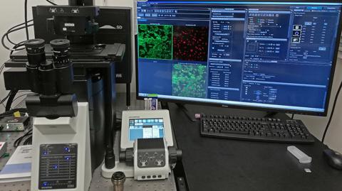
FV3000 Confocal Fluoview
Evident (former Olmpus) Laser scanning Microscope
Lasers: 375 nm, 405 nm, 445nm, 488nm, 514nm, 561nm, 640nm.
Objectives: 1,25x: air, 4x; air, 10x; air, 20x; air, 60x; immersion oil, 100x; silicon oil
- High Sensitivity Multi-Channel Imaging
- Macro to Micro Imaging and Super Resolution
- Increase Productivity with High-Speed Imaging
- Accurate Time-Lapse Imaging
- Superior Objectives
Image
Mica Microhub
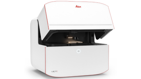
Leica™ Widefield Microhub
- 4 Objectives 1.6x, 10x, 20x, 63x
- 4 LEDs 365 nm, 470 nm, 555 nm, 625 nm
- High resolution Imaging CMOS camera
- Live cell imaging
- Long-term Time Lapse acquisition
- CO2 incubator
- Built-in dark room
- Sample finder
- OneTouch-Auto-Illumination
- AI based analysis
- Integrated Modulation Contrast (IMC)
- Reusable AI models and project parameters
Image
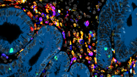
Zellscanner ONE
Zellkraftwerk™ multiplexed imaging Technology
- Highly Multiplexed Tissue Imaging (HMTI) based on cyclic immunofluorescence
- More than 30 markers per cell for cryopreserved and FFPE tissues and more than 100 markers per cell for single cell suspensions
- Antibody stainings can further be combined with mRNA in-situ hybridization (FISH)
- High dynamic range (HDR) imaging technology and true background subtraction for superior image quality
- Microscope performs all steps from sample readjustment, focusing, and image acquisition automatically.
- Pipeline for automated signal quantification developed by us can be used for data analysis
Image
Copyright
Sebastian Jarosch
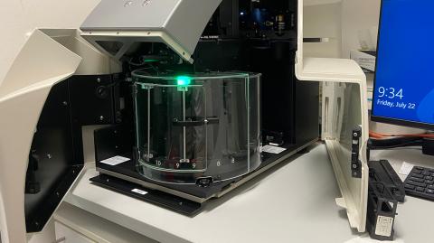
ScanScope Aperio AT2
Leica™ Slide Scanner
Fast, flexible and reliable, it is easy-to-use and consistently delivers high resolution, high quality images.
- Load and capture images of an entire carousel in less than 8 hours
- Sustained high throughput rate of 50 slides/hr with a 20x Objective
- Autoloader with 400-slide capacity
- Smaller file size and faster scanning with skip blank stripes technology
- Horizontal position minimizes risk of coverslips falling off
- Better first time success rates with fewer rescans
- Quiet, with minimal movement of slides in loader
Image
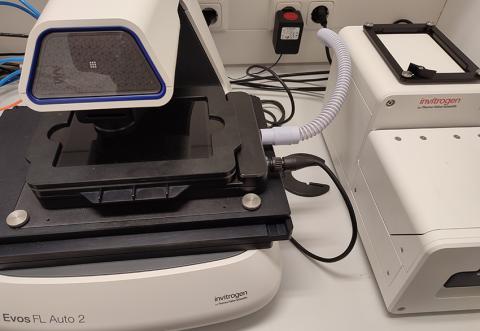
EVOS Cell Imaging System
Invitrogen™
EVOS FL Auto 2
- Speed: scan a 96-well plate in 3 fluorescent channels in less than 5 minutes
- Time-lapse live-cell imaging: option for precise control of temperature, humidity, and gases for normoxic or hypoxic conditions allows a wide range of biological studies under physiological conditions with an onstage incubator
- Area view: move rapidly and seamlessly between low-magnification, single-field mode and high-magnification scan mode to easily define and capture the area of interest
- Automation: time-saving features such as autofocus, rapid stage movement, and automated routines help reduce time to complete experiments, allowing high throughput, high data quality, and improved experimental reproducibility
- Data analysis: advanced software package for quantitative and statistical analysis
Image
IVIS Spectrum
Perkin Elmer™ In Vivo Imaging System
- High-sensitivity in vivo imaging of fluorescence and bioluminescence
- High throughput with 23 cm field of view
- High resolution (to 20 microns) with 3.9 cm field of view
- Twenty eight high efficiency filters spanning 430 – 850 nm
- Supports spectral unmixing applications
- Ideal for distinguishing multiple bioluminescent and fluorescent reporters
- Optical switch in the fluorescence illumination path allows reflection-mode or transmission-mode illumination
- 3D diffuse tomographic reconstruction for both fluorescence and bioluminescence
- Ability import and automatically co-register CT or MRI images yielding a functional and anatomical context for your scientific data.
- NIST traceable absolute calibrations
Image